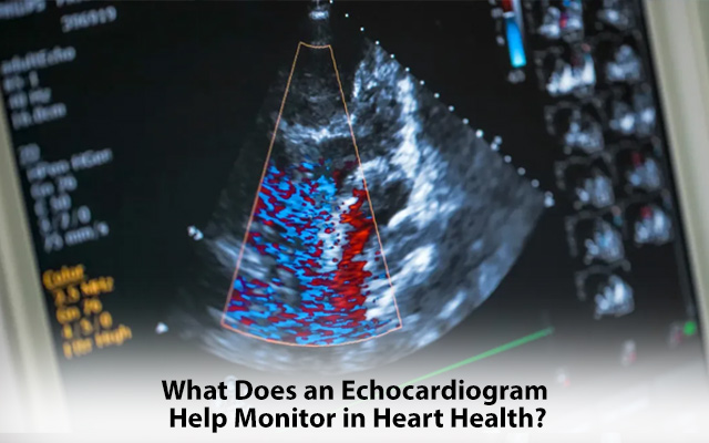
An echocardiogram is a medical test that clearly outlines your heart's structure and function. Using high-frequency sound waves creates live images of the heart, helping doctors assess its health and identify any potential issues.
This non-invasive test is comprehensive, providing a detailed understanding of how well your heart functions, detecting damage, and even spotting congenital issues. In this blog, we will dive into how echo tests in cardiology matter.
Detecting Damage from a Heart Attack
The echo test in cardiology can reveal areas of the heart muscle that have been affected, showing weakened sections that may have reduced ability to pump blood. This information is essential for doctors to assess the extent of damage and plan treatment.
Monitoring How the Heart Pumps Blood
The heart's primary job is to pump blood throughout the body, delivering oxygen and nutrients to vital organs. An echocardiogram helps assess how well the heart is performing this task. It evaluates the heart’s pumping efficiency by looking at the ejection fraction, which measures how much blood the heart pumps with each beat. A lower ejection fraction may indicate heart failure or other heart problems.
Detects Birth Defects Affecting Heart Health
Cardiology echo tests are instrumental in detecting congenital disabilities that may affect the heart. Congenital heart defects are conditions that people are born with, such as holes in the heart walls or improperly formed heart valves.
Identifying Problems with Heart Valves
Your heart has four valves that ensure blood flows in the right direction. If these valves are not functioning correctly, it can lead to various heart issues. An echocardiogram helps identify problems with the heart valves, such as stenosis (narrowing) or regurgitation (leaking). These issues can cause blood to flow inefficiently and strain the heart.
Types of Echocardiograms
There are a few different types of echo tests in cardiology, depending on what information the doctor needs:
- Stress Echocardiogram: This test involves performing an echocardiogram while the heart is under stress, typically induced by exercise or medication. It helps doctors see how the heart performs when it's working harder.
- Transesophageal Echocardiogram (TEE): In this test, a probe is inserted into the oesophagus to get a clearer image of the heart. Since the oesophagus is located close to the heart, this echocardiogram provides more detailed images and can detect problems that may not be visible with a traditional echocardiogram.
Conclusion
An echo test in cardiology is a valuable tool that helps monitor multiple aspects of heart health, from detecting damage from a heart attack to identifying congenital disabilities and heart valve problems. Sound waves to produce detailed heart images help doctors check how well the heart functions and make informed treatment decisions.
Suppose you are concerned about your heart health or experiencing symptoms like chest pain, shortness of breath, or fatigue. In that case, it's important to take control of your health and contact the best cardiology hospital in Kolkata.